 Contact
 Pharmacie
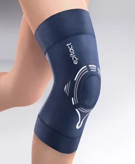
Diagnosing patellofemoral pain syndrome (PFPS) seems easy, but actually, it is quite complicated. Even if this condition hides behind the majority of knee pain complaints, especially in sports medicine, it is necessary to explore any hypothesis.
Indeed, patellofemoral pain syndrome is one of the most common knee conditions. It mainly manifests with pain in the knee joint, between the patella (kneecap) and the femur (thigh bone): the patellofemoral joint.
EPITACT® reviews the diagnosis of patellofemoral pain syndrome: who to consult? How and when is the diagnosis established? Does patellofemoral pain syndrome have a differential diagnosis? What solutions once the diagnosis is confirmed?
Patellofemoral pain syndrome: specialists to consult
To diagnose patellofemoral pain syndrome, do not hesitate to act as soon as the first knee pain appears. Consult a health professional who will identify the cause of your pain. He/She can be a general practitioner, a sports physician or an orthopaedic surgeon.
How to diagnose patellofemoral pain syndrome?
Patellofemoral pain syndrome is diagnosed thanks to several examinations, among which the patient questioning, the physical examination and the radiological assessment, if necessary.
Anamnesis: first step in the diagnosis of patellofemoral pain syndrome
The symptoms of patellofemoral pain syndrome are numerous and vary in intensity but the two main ones are pain and joint instability. Because these symptoms are subjectively perceived by the patient, the practitioner begins the diagnosis with some questions to the patient (anamnesis).
PFPS causes pain in front of the knee, just behind the kneecap. It tends to increase in a sitting position or during repeated flexion/extension movements (e.g.: go up or down the stairs). This questioning is the first step in the diagnosis of patellofemoral pain syndrome or any other disease.
Clinical examination to objectify the pain
Then, it is essential to have a clinical examination to eliminate the main differential diagnoses of patellofemoral pain syndrome. In other words, your GP observes you to assess the normal or abnormal functioning of your knee and the presence of associated conditions(5).
The practitioner generally begins with gait assessment. He subsequently analyses the alignment of lower limbs and your morphotype (for example, genu valgum(1)). To do so, you are standing, in underwear, feet parallel. If necessary, he can ask you to take certain positions like squats to find an eventual muscle weakness (particularly the abductors and quadriceps muscles)(5).
The practitioner can also touch your knee to identify the precise location of pain. Doing patella tests could also be required to assess knee instability or the presence of patellofemoral osteoarthritis.
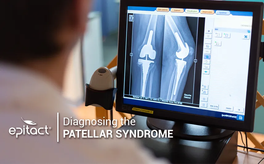
Radiological assessment to confirm the diagnosis of patellofemoral pain syndrome
Diagnosing patellofemoral pain syndrome can be difficult because of the number of possible associated conditions. Therefore, the objective consists in excluding these differential diagnoses of patellofemoral pain syndrome. Since the symptoms of PFPS are not typical of the condition, imaging allows to eliminate some other hypotheses.
Before radiological assessment, the clinical examination aims to reduce the possibilities. For example, a subluxation, a patellofemoral dysplasia or a poor positioning of the kneecap regarding the trochlear groove(2) could be excluded.
Next, medical imaging captures knee X-Rays images from front, profile, weight-bearing and then with the knees bent at 30°(1). MRI can also be required to determine the presence of a cartilage or soft tissues lesion for example(3). The last possible related conditions will be excluded or confirmed: patellar tendinopathy, fracture, Sinding-Larsen-Johansson disease or Osgood-Schlatter disease among others(3, 4).
When to consult in case of knee pain?
If you have knee pain, you should consult as soon as possible to get the diagnosis of patellofemoral pain syndrome Indeed, chronic pain can rapidly become disabling. In the long-term, it can have moral and psychosocial consequences and affect the patient’s quality of life. Every step in the diagnosis of PFPS is crucial, the questioning shouldn’t be neglected.
What solutions are proposed after the diagnosis of PFPS?
After getting the diagnosis of patellofemoral pain syndrome, your general practitioner can prescribe you several treatments, which depend on your symptoms.
Preventive solutions
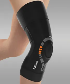
For people subject to patellofemoral pain, preventive solutions exist. EPITACT® has developed two knee support braces. Find out the PHYSIOstrap™ MEDICAL brace* that can be worn daily to secure your knee without limiting your range of motion. Also, discover the PHYSIOstrap™ SPORT brace* that stabilises the kneecap during sports and physical activities.
Curative solutions
Some post-diagnosis solutions are also suggested to relieve pain. Physical therapy sessions with the physical therapist aim to strengthen the muscles around the patella. As a complement, resting and wearing insoles can also be recommended. However, surgery is rarely considered after the diagnosis of patellofemoral pain syndrome.
You’d understand, diagnosing patellofemoral pain syndrome may seem easy but it is actually much more complex. To better understand this condition, learn how to prevent and treat it, and why it is so frequent among sportspersons.
*These solutions are class I medical devices that bear the CE marking under this regulation. Carefully read the instructions before use. Manufacturer: Millet Innovation. 09/2021
For more details about this general and simplified approach, here are further sources:
(1)Tamalet, Bertrand, Pierre Rochcongar, et Goulven Rochcongar. « La fémoro-patellaire : une articulation oubliée ? » Revue du Rhumatisme Monographies, Pathologies du genou - Première partie, 83, no 2 (1 avril 2016): 71 77. https://doi.org/10.1016/j.monrhu.2016.01.005.
(2)Goux, P Le. « Démembrement clinique et approche thérapeutique des syndromes rotuliens - Clinical analysis and treatment of the patellofemoral syndromes ». Mise au point, La Lettre du Rhumatologue, no 315 (2005): 5.
(3)Fournier, Dr Yann. « Le Syndrome Douloureux Rotulien ». Centre orthopédique Santy, 2015. http://orthopedie-lyon.fr/wp-content/uploads/2012/02/DIU-SPORT_-LYON_-SYNDROME-DOULOUREUX-ROTULIEN_DR-FOURNIER-min.pdf.
(4)Baptiste Claudon. Phénomène d’adaptation cinétique lors de la marche du patient présentant un syndrome femoro-patellaire douloureux : mise en évidence et réversibilité sous traitement de rééducation. Sciences du Vivant [q-bio]. 2010. hal-01733971
(5)Willy RW, Hoglund LT, Barton CJ, et al. Patellofemoral pain: clinical practice guidelines linked to the International Classification of Functioning, Disability, and Health from the Academy of Orthopaedic Physical Therapy of the American Physical Therapy Association. J Orthop Sports Phys Ther. 2019;49(9):CPG1–CPG95. doi:10.2519/jospt.2019.0302.